WHAT IS RETINITIS PIGMENTOSA?
SYMPTOMS
CURRENTLY AVAILABLE TREATMENTS
CASE STUDIES
CONTACT FOR MORE INFORMATION
Retinitis Pigmentosa is the name for a group of genetic diseases that are the leading cause of inherited blindness. It affects an estimate 1.5 million people worldwide.
People with retinitis pigmentosa lose their vision because light-sensitive retinal photoreceptor cells in their eyes gradually die.
Patients – some as young as 17 years – typically experience a progressive loss of vision, “tunnel” vision, and finally legal blindness, with devastating consequences for their future lives.
There is no known cure.
What Causes Retinitis Pigmentosa?
Vision loss in retinitis pigmentosa happens because light-sensitive “photoreceptor” cells become damaged and die as a result of harmful changes – known as ‘mutations’ – in more than 100 different genes.
Genes determine the type and amount of proteins in cells. Because of these harmful mutations, retinal photoreceptors or their supporting cells make defective proteins – or they may have too much or too little of a particular protein, leading to abnormal function – and eventually, the death of retinal photoreceptors.

Left: Normal eye. Right: Eye with the typical pigmentation seen in Retinitis Pigmentosa (Images courtesy Dr. Erika Lygerou-Meropi, OMMA Ophthalmological Clinic, Athens)
These genetic mutations are inherited. In other words, they can be transferred from a parent to a child either in an autosomal dominant, autosomal recessive, X-linked, or maternal (mitochondrial) manner depending on the specific gene mutations present in the parent.
On average, 50% of patients with retinitis pigmentosa will have a history of at least one other family member being affected – while the remaining 50% may not have a family history at all.
So if you have a family member that has already been diagnosed with retinitis pigmentosa, we strongly recommend that you and other members of your family get your eyes checked immediately by an ophthalmologist who is specially trained to detect retinal diseases.
Retinitis pigmentosa is typically diagnosed in adolescents and young adults, some of whom are only 16-17 years old.
Symptoms such as night blindness, loss of peripheral vision, cataracts, and loss of central vision may manifest as early as childhood and are usually full-blown by early adulthood. In later stages, both color perception and central vision may also be lost.
Finally, these unfortunate people end up experiencing “tunnel vision” with a central visual field of less than 20 degrees in diameter, typically becoming legally blind by the age of 40-50 years.
If you have been diagnosed with retinitis pigmentosa, you are likely to experience the following symptoms:
- Night Blindness and Progressive Loss of “Peripheral” Vision – along with these, you may also experience difficulties adapting between well-lit and dark surroundings.
- Diminished Color Vision – over time, both your range of vision and color vision are likely to become worse. Unfortunately, these symptoms usually progress at the same rate in both eyes.
- Tunnel Vision – as retinitis pigmentosa progresses, you will develop “tunnel vision” – along with difficulty reading, driving and walking without assistance as well as recognizing faces and objects.
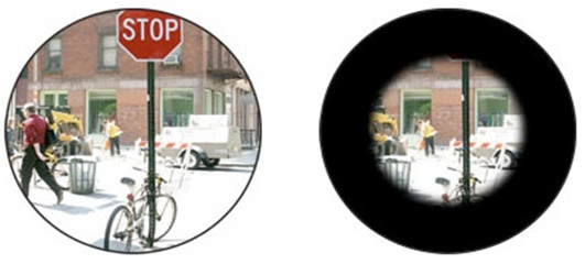
Left: Normal vision. Right: “Tunnel” vision seen in Retinitis Pigmentosa (Images courtesy Dr. Erika Lygerou-Meropi, OMMA Ophthalmological Clinic, Athens)
- Photophobia – you may also experience sensitivity to bright light.
- Blindness – eventually you are likely to lose most of your eyesight, becoming legally blind.
Although this is the general experience of most patients with retinitis pigmentosa, the exact symptoms and their progression may differ from person to person and even among affected members of the same family.
For instance, some patients retain central vision and a restricted field of vision well into their 50s, while others experience significant vision loss in their teens or early adulthood.
Both eyes are usually affected in a similar way.
There are presently no proven cures for retinitis pigmentosa. In fact, even efforts to slow down the progression of this inherited retinal disorder have not been very successful.
A 2014 review of 3 clinical studies conducted in the U.S. and Canada found that administering high‐dose vitamin A and fish oils does not significantly slow or prevent vision loss in patients with retinitis pigmentosa.
There is some speculation that treatment with neuroprotective agents such as valproic acid, ciliary neurotrophic factor (CNF), and calcium channel inhibitors – or supplementation with vitamin A, lutein, synthetic retinoids, and decosahexaenoic acid may slow down progression of visual field loss and positively influence eye electrophysiological parameters. However, the potential adverse effects of giving these compounds over the long-term are not well known.
Microelectronic chip technologies including retinal prosthesis, artificial vision, retinal chip, and bionic eye are currently being tested in patients with retinitis pigmentosa, with varying degrees of success.
There is no question that these aids have removed many barriers to education and employment and made patients with retinitis pigmentosa more capable of leading independent lives.
However, no drug or treatment available today can halt the progressive loss of vision seen in patients with Retinitis Pigmentosa, much less restore lost vision.
Until today.
Incredibly, the breakthrough new treatment Rejuven-Eyes Bio-Therapy has been clinically proven to slow down vision loss – even restore lost vision – in patients with Retinitis Pigmentosa.
No other vision loss therapy can do this.
BENEFITS OF REJUVEN-EYES BIO-THERAPY
- Stops Ongoing Vision Loss After Just One Treatment Course
This innovative therapy stopped ongoing vision loss quickly and painlessly in patients suffering from retinitis pigmentosa after just one course of treatment lasting 20 days.
- Restores Lost Vision
A significant proportion of patients reported a noticeable improvement in their eyesight on treatment with Rejuven-Eyes Bio-Therapy.
Yes, it’s true – they regained some of their lost vision.
- Is Clinically Proven to be 100% Safe
Not a single patient reported any side effects or adverse reactions. In other words, clinical data based on patient outcomes clearly confirms that Rejuven-Eyes Bio-Therapy is fast-acting, powerfully effective and 100% safe.
- Does Not Involve Intraocular Injections
This novel therapy is NOT injected directly into the eyes, but into the muscle like a flu shot. In fact, patients report that this treatment is comfortable and completely painless.
Patients with retinitis pigmentosa experience a gradual loss of vision because the two types of photoreceptor cells in the retina of their eyes – known as rod and cone photoreceptor cells – die.

Human eye: Retina in detail, with rod and cone photoreceptor cells (From ASU School of Life Sciences website https://askabiologist.asu.edu/)
Rod cells are present throughout the retina, except for the very center, and they are needed mainly for night vision.
Cone cells are also present throughout the retina. However, they are much more concentrated in the central region of the retina, known as the macula. Cone cells are necessary for color vision and “sharp” central vision – which helps us to read, see details of faces and colors etc.
In retinitis pigmentosa, first the rod cells and later the cone cells stop working.
This is why night and peripheral vision are affected first, while many patients still retain useful color and sharp central vision well into middle age.
As their peripheral vision becomes increasingly compromised, patients with retinitis pigmentosa will experience progressive "tunnel vision”. Eventually, as more and more of their cone cells die, they will inevitably progress to legal or complete blindness.
How does Rejuven-Eyes Bio-Therapy help to prevent ongoing vision loss, even restore lost vision in patients with Retinitis Pigmentosa?
The synergistic formulation of small but therapeutically potent “retinoprotective” peptides in Rejuven-Eyes Bio-Therapy halt the ongoing damage of both rod and cone photoreceptor cells. This innovative new therapy is also believed to trigger the formation of new, healthy photoreceptor cells!
For instance, one of the main components of Rejuven-Eyes Bio-Therapy known as Retinalamin has been shown to trigger formation of new retinal cells, significantly improving visual acuity and expanding the field of vision in patients. Another component known as Epitalon has been shown to improve visual functions in up to 90% of patients with the symptoms of pigmented retinal degeneration that are typically seen in retinitis pigmentosa and other related eye conditions.
Let’s consider individual case studies of patients with retinitis pigmentosa treated with Rejuven-Eyes Bio-Therapy.
CASE STUDY 1 – Male, 17 years old, OMMA Ophthalmological Institute, Athens
Clinical Observations before treatment
Visual Acuity in Left Eye – 20/32; Visual Acuity in Right Eye – 20/32
Note: “Visual acuity” means clarity or sharpness of vision and is measured separately for each eye. 20/20 vision is normal vision. If you have 20/20 vision, you can see objects in clear detail at 20 feet which the average person can see at that distance.
If you have 20/32 vision, then you need to be as close as 20 feet to clearly see the objects that an average person with normal eyesight can see from 32 feet away.
Clinical Observations after two courses of treatment with Rejuven-Eyes Bio-Therapy
- Improvement in Left Eye – significant expansion of central photosensitive area; in other words, after 2 courses of treatment more photoreceptor cells in the retina of this patient’s left eye are responding to light, helping him to see.
Visual Acuity improved to 20/29. - Improvement in Right Eye – significant expansion of central photosensitive area; after 2 courses of treatment, more photoreceptor cells in the retina of this patient’s right eye are responding to light, helping him to see.
Visual Acuity improved to 20/29.
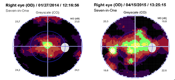
Left image: Central photosensitive area in right eye of 17-year-old male patient (Case Study 1) before treatment.
Right image: Central photosensitive area in right eye of this patient expanded significantly after two courses of treatment with Rejuven-Eyes Bio-Therapy.

Left image: Central photosensitive area in left eye of 17-year-old male patient (Case Study 1) before treatment.
Right image: Central photosensitive area in left eye of this patient also expanded significantly after two courses of treatment with Rejuven-Eyes Bio-Therapy.
CASE STUDY 2 – Male, 33 years old, OMMA Ophthalmological Institute, Athens
Clinical Observations before treatment
Visual Acuity in Left Eye – 20/67; Visual Acuity in Right Eye – 20/29
Clinical Observations after 2 courses of Rejuven-Eyes Bio-Therapy
- Improvement in Left Eye – Visual Acuity improved to 20/40.
- Improvement in Right Eye – Visual Acuity improved to 20/22.
CASE STUDY 3 – Female, 39 years old, OMMA Ophthalmological Institute, Athens
Clinical Observations before treatment
Visual Acuity in Left Eye – 20/40; Visual Acuity in Right Eye – 20/40
Clinical Observations after treatment with Rejuven-Eyes Bio-Therapy
- Stabilization of Left Eye. Visual Acuity remained 20/40.
- Improvement in Right Eye – significant expansion of “central photosensitive area”; more photoreceptor cells in the retina of this patient’s right eye are responding to light and helping her to see. Visual Acuity remained 20/40.
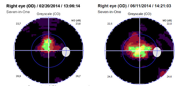
Left image: Central photosensitive area in right eye of 39-year-old female patient (Case Study 3) before treatment.
Right image: Central photosensitive area in right eye of this patient expanded after one course of treatment with Rejuven-Eyes Bio-Therapy.

Left image: Central photosensitive area in left eye of 39-year-old female patient (Case Study 3) before treatment.
Right image: Central photosensitive area in right eye of this patient stabilized after one course of treatment with Rejuven-Eyes Bio-Therapy.
CASE STUDY 4 – Male, 54 years old, St. Petersburg Institute of Bioregulation and Gerontology, Russia
Clinical Observations before treatment
Visual Acuity in Left Eye – 20/2857; Visual Acuity in Right Eye – 20/4000
Clinical Observations after treatment with 4 courses of Rejuven-Eyes Bio-Therapy
- Improvement in Left Eye. Visual Acuity improved to 20/1000.
- Improvement in Right Eye – Visual Acuity improved to 20/1000.
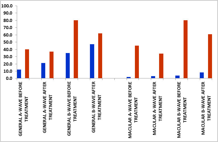
Electroretinographic (ERG) Data, CASE STUDY 4
The electroretinogram is a diagnostic test that measures electrical activity generated by neuronal and non-neuronal cells in the retina in response to a light stimulus. ERGs are obtained using electrodes embedded in a corneal contact lens, which measure a summation of retinal electrical activity at the corneal surface. They can provide important diagnostic information on a variety of retinal disorders and can also be used to monitor eye disease progression.
The components of a typical ERG include:
A-waves: derived from the cones and rods of the outer photoreceptor layers.
B-waves: derived from the inner retina, this wave is the most common component of the ERG used in clinical and experimental analysis of human retinal function.
GRAPH 1: Changes in amplitude (MCV, blue columns) and latency (in MS, brown columns) of A- and B- waves before and after 4 courses of treatment with Rejuven-Eyes Bio-Therapy in the left eye of a 54-year-old patient with Retinitis Pigmentosa.
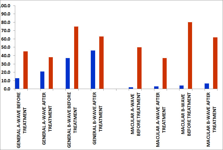
Results: In both the overall retina and the macula of the left eye, amplitude of both A- and B-waves (blue columns) increases after treatment with Rejuven-Eyes Bio-Therapy, while latency (brown columns) decreases proportionately.
GRAPH 2: Changes in amplitude (MCV, blue columns) and latency (in MS, brown columns) of A- and B- waves before and after 4 courses of treatment with Rejuven-Eyes Bio-Therapy in the right eye of a 54-year-old patient with Retinitis Pigmentosa.
Results: In both the overall retina and the macula of the right eye, amplitude of both A- and B-waves (blue columns) increases after treatment with Rejuven-Eyes Bio-Therapy, while latency (brown columns) decreases proportionately.
In other words, treatment with Rejuven-Eyes Bio-Therapy increased functional electrical activity of both cone and rod photoreceptor cells in the inner as well as outer layers of the retina in both eyes of this patient. This corresponds to the increase in visual acuity seen in both eyes.
The harsh reality is that if you have been diagnosed with Retinitis Pigmentosa, no drug or treatment available today can save your vision.
However, there’s no need to lose hope.
Rejuven-Eyes Bio-Therapy has been shown to –
- Quickly and painlessly stop progression of vision loss in patients with Retinitis Pigmentosa after just 20 days of treatment!
- Even restore lost vision in a significant proportion of patients
- Without causing any side effects or adverse reactions in a single patient.
So if you have been diagnosed with Retinitis Pigmentosa, or know someone who does –
We strongly recommend that you get in touch with us right away to learn how you can prevent further damage to your precious vision – and perhaps even recover some of your lost vision!